Faradic and Galvanic Machines · Volume 3
Duchenne and the Living Muscle
How Guillaume-Benjamin-Amand Duchenne de Boulogne turned the induction coil into an instrument for reading the body — mapping which muscles a current could twitch or tetanize, arguing the faradic current's case against the galvanic, and helping found the discipline of electrotherapy. The pioneers around him, and the portable medical-battery trade that followed.
Contents
3.1 About this volume
Vol 1 — The Two Currents and the Medical Battery established the two workhorse currents of nineteenth-century electrotherapy — the steady galvanic direct current of a chemical cell, and the pulsed, induced faradic current struck from an interrupted induction coil — and the fitted wooden “medical battery” that delivered them. Vol 2 — What the Current Does to Tissue took those two currents down to the tissue, separating the genuine electrochemical effects of galvanic DC at the electrode from the depolarisation of motor nerves by faradic pulses.
This volume is about the person who did more than anyone to turn that apparatus from a curiosity into a clinical method, and about the community of workers around him. Guillaume-Benjamin-Amand Duchenne de Boulogne is routinely called the “father of electrotherapy,” a phrase the Science Museum Group itself uses of the man whose 1849 induction machine it holds. The label is a convenience — the field had many parents — but Duchenne earns a central place in it, because he was the first to use the induced current systematically not merely to treat but to investigate: to make a living muscle contract on command and, from the contraction, to read the muscle’s function and the state of its nerve.
Two disciplines are careful to note throughout. First, the dates and priority claims in this field are contested and were sometimes tidied up after the fact; where a claim rests on a secondary source or a single museum caption, this volume says so rather than manufacture precision. Second, nothing here is a procedure. This is the history of how a method of examination was built, not an account anyone could act on.
3.2 The man and the making of a field
Duchenne was born in Boulogne-sur-Mer on 17 September 1806, the son of a sea captain, and died in Paris in 1875. (One museum record gives his birth year as 1809; the overwhelming weight of biographical sources puts it at 1806, and this volume follows them.) He trained in medicine in Paris under the leading clinicians of the day, practised for a time back in Boulogne, and in 1842 returned to Paris, where — by his own account, “working with an ardour that has rarely been equalled” — he spent the rest of his life on a single obsession: what a controlled electric current could reveal about muscle and nerve.
He did not invent medical electricity, and he was not the first to push a current into a muscle. From about 1835 Duchenne experimented with électropuncture, a technique introduced by François Magendie and Jean-Baptiste Sarlandière in the 1820s in which a fine needle electrode was driven through the skin to shock the muscle beneath. Duchenne’s decisive practical insight was that he did not need to break the skin at all: a current delivered through moistened pad electrodes pressed on the surface could be steered to a chosen muscle far less painfully. That move — from the needle to the wet surface electrode — is the foundation of everything that follows, and it is why his machines carry cloth-covered electrode handles rather than lancets.
3.3 Localised electrisation: reading the living muscle
Duchenne called his method “électrisation localisée” — localised electrisation — and gave it its definitive statement in the treatise De l’électrisation localisée et de son application à la pathologie et à la thérapeutique, first published in 1855 and expanded through further editions (a third by 1872). The idea was simple and radical: by placing surface electrodes precisely and choosing the current carefully, he could make one muscle, or one head of a muscle, contract in isolation while its neighbours stayed still. Watching the movement that resulted, he could deduce what that muscle actually did.
The interrupted faradic current was the right tool for this because its behaviour could be varied. A single make-and-break of the interrupter gives a single pulse and a single twitch; run the interrupter fast enough and the twitches fuse into a sustained, smooth tetanic contraction — the muscle held taut for as long as the current flows. By changing how fast the coil’s interrupter buzzed, and how strong the induced pulse was, Duchenne could dial a muscle’s response from a flick to a locked contraction, and hold it there long enough to observe and (in his later work) photograph it. The physiology underneath this — why a fast enough train of nerve depolarisations sums into tetanus — is Vol 2’s subject; here the point is that Duchenne turned that graded response into an examining tool.
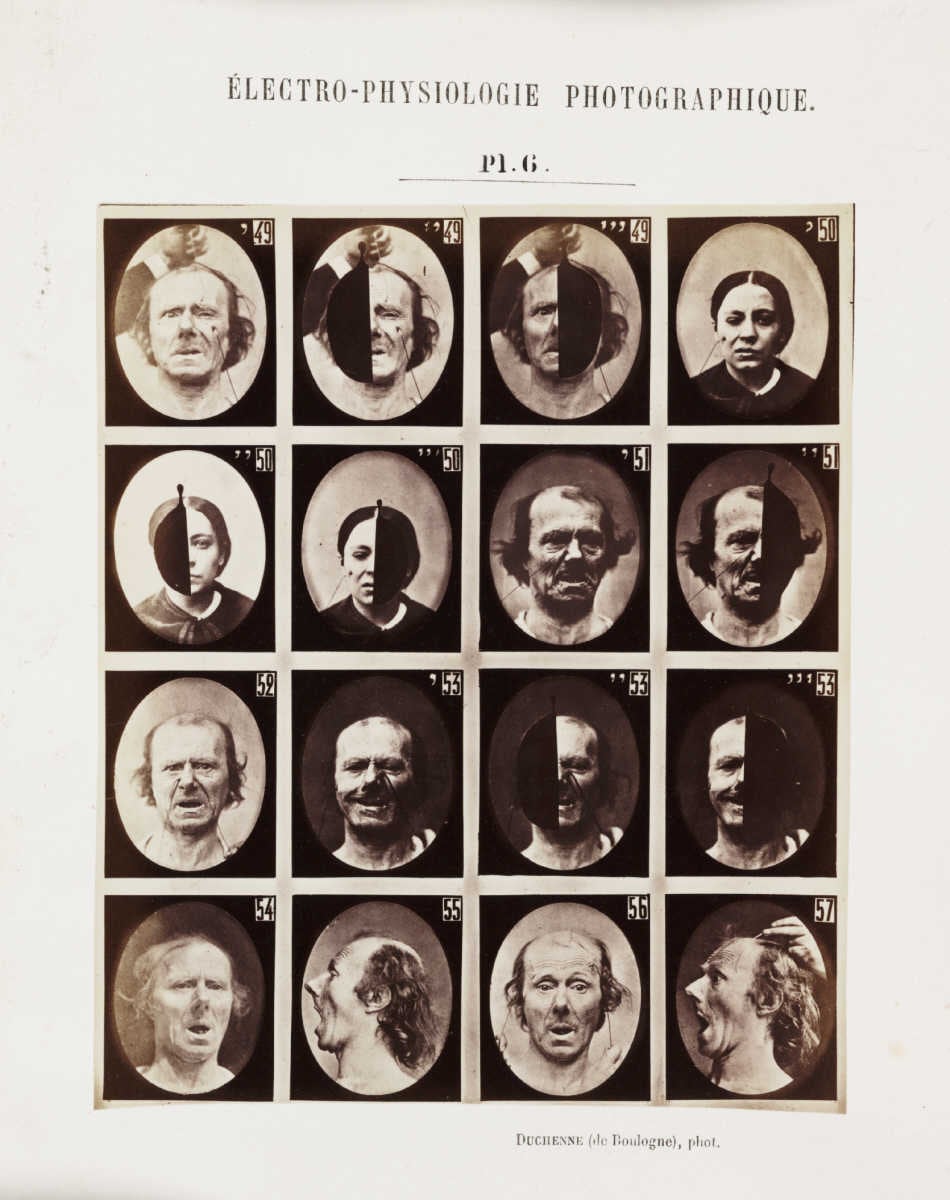
Out of thousands of such stimulations he built what amounted to an atlas of the living musculature, showing that ordinary movements are not the work of single muscles but of coordinated teams of agonists and antagonists. He carried the method into diagnosis, using the muscle’s electrical response — whether it still answered a faradic pulse at all, and how — as a sign of the health of the nerve supplying it. In doing so he described neuromuscular disorders that still bear his association: the muscular dystrophy that carries his name, the wasting of progressive muscular atrophy (the Aran–Duchenne form), and the locomotor ataxia of tabes dorsalis. His most famous single application was the least therapeutic: in Mécanisme de la physionomie humaine (1862) he faradised individual facial muscles to reproduce human expressions on command, photographing the results — work Charles Darwin drew on directly for The Expression of the Emotions in Man and Animals (1872).
3.4 Faradic versus galvanic: the argument and its limits
Duchenne tried both of the era’s currents and came down firmly for the induced one. He named it “faradic” after Michael Faraday, who had discovered electromagnetic induction in 1831, and around 1855–56 he argued that the induced (interrupted, roughly alternating) current was superior to the steady galvanic current for the specific purpose of triggering muscle contraction. The reasoning that comes down to us is practical and physiological: to make a muscle contract with steady direct current you must push the current hard, and at that strength galvanic DC produces a “warming,” irritant, blistering effect at the skin under the electrode — the electrochemical action described in Vol 2 — whereas a brief faradic pulse fires the motor nerve and gets the contraction without cooking the skin.
It is worth being precise about what this claim was and was not. The frequently repeated statement that Duchenne “announced in 1856 that alternating was superior to direct current” for triggering contraction rests largely on secondary summaries rather than a quoted line of his; the substance is sound and consistent with his 1855 treatise, but the tidy one-sentence, one-year form should be read as a paraphrase of a position he built up over several years, not a dated pronouncement. Treat the “1855–56” anchor as approximately right and deliberately hedged.
And Duchenne’s preference did not settle the matter — which is the historically honest point. The steady galvanic current has real effects the faradic current does not (the electrochemical, iontophoretic, and electrolytic actions of Vol 2), and it kept its own school of advocates. His methodology and conclusions drew him into a sharp, extended debate with the German physician Robert Remak (1815–1865), who championed galvanotherapy — the therapeutic use of steady DC — and who in 1858 drew attention to the “motor points” where a nerve enters its muscle. The nineteenth century did not choose faradic over galvanic so much as learn to use each for what it does best: faradic to drive contraction and test the nerve–muscle response, galvanic for its slower chemical and (as later framed) sedative effects. Both survive in modern physiotherapy, exactly as Vol 2 sets out.
3.5 The pioneers around him
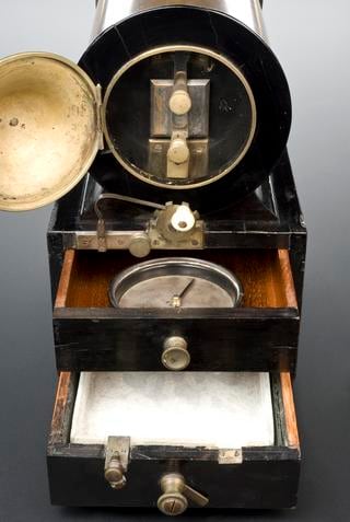
Calling Duchenne the “father” of the field flattens a crowded genealogy, and the sober account names the others. The physics was not his: Faraday’s 1831 discovery of induction supplied the effect, and the instrument-makers turned it into hardware — Heinrich Daniel Ruhmkorff commercialised the large induction coil (patented 1851, from his Paris workshop), the object that gave “Ruhmkorff coil” to the language. A useful chronological caution follows from that date: Duchenne’s induction machine is dated 1849 and was built by the Paris surgical-instrument maker Charrière, so it slightly predates the classic Ruhmkorff coil and is better described as an early medical induction apparatus than as a Ruhmkorff proper. Later Duchenne-associated machines took other forms again — the Museo Galileo holds a portable magneto-electric (“magneto-faradic double-current”) apparatus of about 1870 by the Paris maker Deleuil, in which a hand-cranked armature spinning past a permanent magnet, rather than an interrupted battery-and-coil, generated the current.
The electrophysiology had other founders too. Carlo Matteucci and, above all, Emil du Bois-Reymond established that nerve and muscle themselves generate and respond to electrical signals, the science that made Duchenne’s diagnostic readings interpretable. And the diagnostic method Duchenne began was systematised by the German school after him: Wilhelm Erb (1840–1921), working in Leipzig and Heidelberg, built electrodiagnosis into a formal clinical discipline resting on both faradic and galvanic testing, and described the reaction of degeneration (Entartungsreaktion) — the tell-tale change in how a denervated muscle answers the two currents, which let a physician distinguish a nerve lesion from a muscle one. Duchenne is the father in the sense that he moved first and moved the field’s centre of gravity from spectacle to method; Remak, Erb, du Bois-Reymond, Matteucci, and the instrument-makers are the reason it became a durable clinical tradition rather than one man’s technique.
3.6 From the physician’s machine to the portable medical battery
Duchenne’s own machines were serious clinical instruments — the Charrière induction apparatus, the Deleuil magneto — kept and used by a physician who understood exactly what a given current did. But the same core hardware, an induction coil with an interrupter and a set of electrode handles, was eminently portable and eminently saleable. Through the second half of the century a trade in cased “medical batteries” grew up on both sides of the Atlantic, packaging coil, cells, interrupter, and electrodes into the fitted mahogany and walnut boxes that are the collector’s quarry today — the objects Vol 5 — Collecting the Medical Battery teaches how to read and date.
That portability cut both ways, and it is the hinge into the rest of this dive. In a physician’s hands the cased faradic set was a legitimate instrument for muscle re-education and for testing nerve injury; in a canvasser’s hands the very same box became a cure-all sold for “nervous exhaustion,” lost vigour, and impotence, and its shock-coil turned up as the fairground grip-test shock box. That split — genuine physical medicine on one side, carnival and cure-all marketing on the other — is Vol 4 — Legitimate Medicine and Carnival’s to work out, and it runs straight into the Quack wing: the galvanic “electric belts” of the Electropathic Belts dive borrowed Duchenne’s respectable galvanic current for wearable appliances that mostly did nothing, and the high-frequency apparatus a hospital called d’Arsonvalization was sold door-to-door as the Violet Ray Wands handset. The sober high-frequency line that grew out of this same tradition is picked up in the High Frequency Currents dive of this wing.
3.7 Where this volume hands off
Duchenne gives the dive its human centre: the man who took the two currents of Vol 1 and the tissue effects of Vol 2 and made them into a way of reading the body, and the pioneers around him who turned that into a discipline. The next volume follows the same hardware as it splits in two — into the legitimate medicine of diagnosis, iontophoresis, electrolysis, and muscle re-education, and into the cased “medical battery” sold for vigour and the arcade shock box — in Vol 4 — Legitimate Medicine and Carnival. The physical afterlife of these machines, and how a collector identifies and dates a fitted case, closes the dive in Vol 5 — Collecting the Medical Battery.
Sources
- Duchenne Medical Induction Machine, Paris, 1849 — Science Museum Group Collection (object co140173) — the surviving 1849 machine: maker Charrière, Paris; ebony base with brass fittings; induction coil; “localised electrization” with cloth-covered electrodes; the “father of electrotherapy” phrasing (and the outlier 1809 birth year, corrected here).
- Duchenne’s Medical Magneto-Electrical Machine — Museo Galileo (Inv. 456) — a c.1870 portable magneto-faradic double-current apparatus by Deleuil, Paris; current generated by a hand-cranked armature past a permanent magnet; electrode set — used here for the machine-form evolution and to distinguish magneto from induction-coil designs.
- Duchenne de Boulogne — Hektoen International (2019) — Duchenne’s 1842 move to Paris and start of faradic work on skeletal muscle; électrisation localisée as a mapping method (coordinated agonists/antagonists); the 1855 treatise and its editions; the 1862 Mécanisme de la physionomie humaine and Darwin’s use of it.
- Guillaume Duchenne de Boulogne — Wikipedia (leads, cross-checked) — birth 17 September 1806, Boulogne-sur-Mer; death 1875; 1835 électropuncture (after Magendie and Sarlandière) and the shift to moistened surface electrodes; “faradic” named for Faraday (induction, 1831); the 1855 title and the associated neuromuscular disorders.
- Electrotherapy — Wikipedia (leads, cross-checked) — the ~1856 statement that induced (“alternating”) current was superior to direct current for triggering muscle contraction, and the “warming”/blistering irritant effect of DC at contraction-strength — treated here as a hedged paraphrase, not a dated quotation.
- The Origins of Neuromuscular Electrodiagnosis, 1800–1950 — European Neurology (Karger) — the wider pioneer genealogy: Duchenne’s debate with Robert Remak (galvanotherapy; 1858 motor-points work), and Wilhelm Erb’s systematisation of electrodiagnosis and the reaction of degeneration (Entartungsreaktion).
- Heinrich Daniel Ruhmkorff — Wikipedia (leads, cross-checked) — the induction coil patented 1851 from Ruhmkorff’s Paris workshop, used to establish that Duchenne’s 1849 Charrière machine predates the classic Ruhmkorff coil.
Comments (0)