Electrotherapy Devices · Volume 5
Legacy, Identifying and Collecting
The honest verdict on medical electricity is neither “it was all quackery” nor “the Victorians were right all along.” It is stranger and more interesting than either. The nineteenth century genuinely glimpsed real physiology — that nerve and muscle run on electrical signals, that current can make a muscle twitch, that high-frequency current heats tissue without a violent shock — and then sold those glimpses as cures for everything from paralysis to “nervous exhaustion.” The cure-all claims died. The physiology did not. It matured, quietly and unglamorously, into some of the most consequential technology in twentieth-century medicine: the pace of a stopped heart restarted across a chest, a chronic-pain patient reaching for a battery pack instead of surgery, a diagnostic needle listening to a single motor unit fire. This volume traces that crossover, then turns to the practical business of identifying, dating, and — above all — not powering up the antique apparatus these ideas left behind.
This is the closing volume of the History dive. It cross-links the five device dives — Static Electricity Machines, Faradic and Galvanic Machines, High-Frequency Currents, Diathermy Machines, and Early Electroconvulsive Therapy — rather than repeating their detail.
5.1 Pain, Reframed: TENS and Electroanalgesia
The oldest promise of medical electricity was pain relief, and it is the one that came back most decisively — but only after the reason it sometimes worked was finally explained. In 1965 Ronald Melzack and Patrick Wall published the gate control theory of pain, arguing that stimulating large sensory (Aβ) fibres could “close a gate” in the spinal dorsal horn and inhibit the transmission of pain signals carried by small nociceptive (C and Aδ) fibres. For the first time there was a physiological rationale for why buzzing the skin might dull pain, rather than the vague “nervous tonic” language of the electrotherapy catalogues.
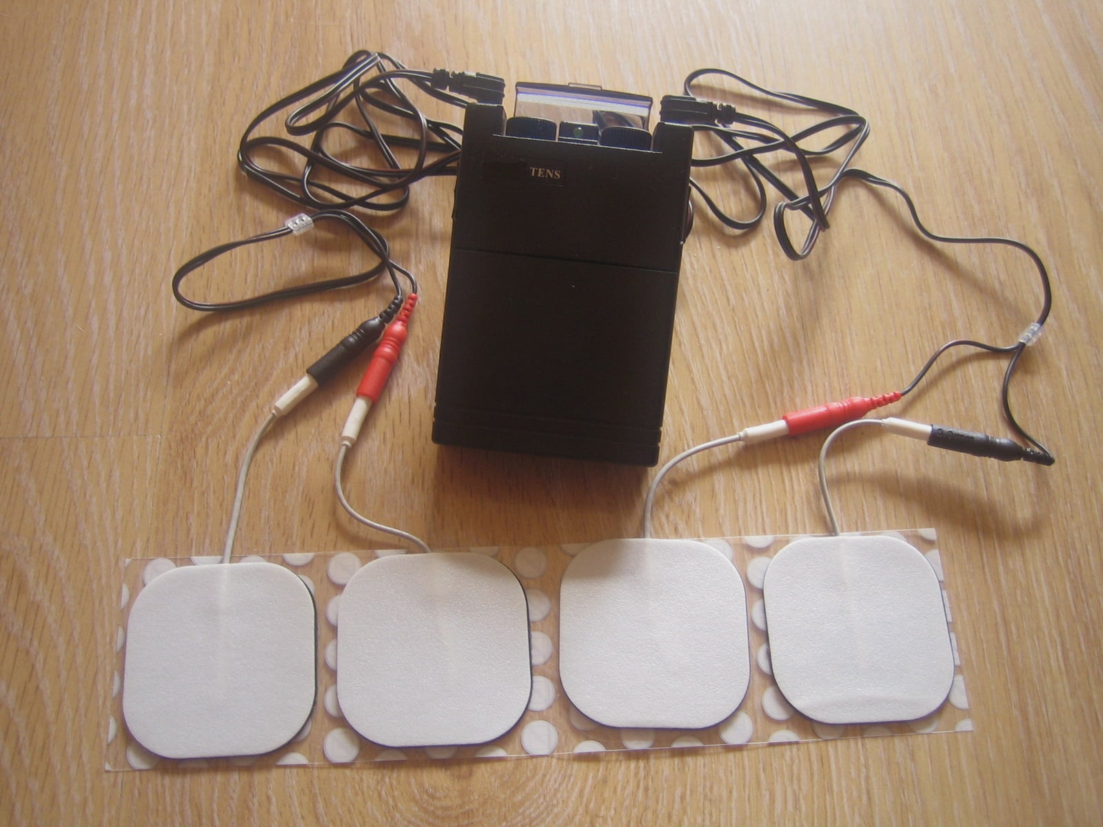
The American neurosurgeon C. Norman Shealy turned the theory into hardware. From 1967 he developed a surgically implanted dorsal-column stimulator, and used a portable skin-surface stimulator as a screening tool — to predict which chronic-pain patients would benefit from the implant. The twist that made history: large numbers of patients found the external device relieved their pain well enough that they declined the implant altogether. That external screening device is the direct ancestor of modern TENS (transcutaneous electrical nerve stimulation), now a mass-market, over-the-counter pain therapy. The through-line to this wing is exact — a faradic “medical battery” (see Faradic and Galvanic Machines) also passed pulsed current through skin electrodes for pain. The difference is that TENS rests on a tested mechanism and makes modest, bounded claims, where its Victorian ancestor promised to cure the disease itself.
5.2 The Heart: Defibrillation and Cardiac Pacing
Nowhere did the crossover matter more than the heart, and nowhere is the lineage from the shock box to the clinic clearer. Once physiologists understood that the heartbeat is an electrical event, two questions followed: could you restart a fibrillating heart with a shock, and could you pace one that beat too slowly?
Defibrillation arrived in stages. Paul Zoll demonstrated successful external (closed-chest) defibrillation in humans in 1956, reversing ventricular fibrillation with a shock delivered through the intact chest wall — cited at up to around 750 volts of alternating current. William Kouwenhoven at Johns Hopkins built an early closed-chest AC defibrillator around 1957 delivering roughly 480-volt shocks, but the machine weighed over 250 pounds and was anything but portable. The decisive refinement came from Bernard Lown: in a 1962 JAMA paper, Lown, Amarasingham, and Neuman reported treating ventricular tachycardia with a synchronised direct-current (DC) shock discharged from a capacitor — the “Lown waveform” — after concluding that AC caused more arrhythmias and tissue damage. DC cardioversion is essentially the technique still in use today, and Kouwenhoven’s team’s parallel work on closed-chest cardiac massage helped establish modern CPR.
Cardiac pacing ran on a parallel track. Zoll had already demonstrated the first successful external cardiac pacemaker in 1952, but it tethered the patient to a mains-powered console at the bedside. The leap to a fully implantable pacemaker came in Sweden in 1958 (the Senning–Elmqvist device implanted in patient Arne Larsson). In the United States, engineer Wilson Greatbatch and surgeon William Chardack implanted their transistorised, mercury-zinc-battery pacemaker at the Buffalo Veterans Hospital in 1960 — a device Greatbatch famously refined on a shoestring in his backyard workshop. That the Bakken Museum, the world’s only museum devoted to medical electricity, displays pacemakers alongside eighteenth-century static machines is no accident: they are the same intellectual project, two centuries apart. It is worth stating plainly against the quack record — a violet-ray wand (see High-Frequency Currents) never restarted a heart, and never could. The defibrillator did, because it was built on measured cardiac electrophysiology rather than on marketing.
5.3 Deep Heat Grows Up: Diathermy to Modern Physiotherapy
The one nineteenth-century discovery with a nearly uncontested clinical afterlife is that high-frequency current heats tissue without causing the tetanic, painful muscle contraction of ordinary shock. That observation, made in the d’Arsonval era, led straight to diathermy — deliberate deep heating of muscle and joint for physical medicine — a term generally credited to Karl Nagelschmidt around 1908. Early long-wave, spark-gap diathermy gave way in the 1920s to vacuum-tube short-wave diathermy, which by international agreement was allocated the industrial-scientific-medical band at 27.12 MHz, with microwave diathermy following later. Energy was coupled through the body by capacitive plates or inductive coils rather than direct-contact electrodes, reducing burns.
This is the legitimate maturation of the technology traced in High-Frequency Currents and Diathermy Machines. Modern physiotherapy still uses these principles in refined, regulated forms — shortwave and, more commonly today, therapeutic ultrasound — for pain, stiffness, and circulation. The field also taught the profession real caution: deep burns, hazards around metal implants, and later concern over radio-frequency exposure drove much of the electrical-safety practice that now governs medical devices. The overreach — mid-century claims that diathermy could cure infection or cancer — quietly fell away, but the heating chair in the physiotherapy department is a direct descendant of the hospital d’Arsonval apparatus.
5.4 Reading the Body’s Own Currents: EMG and Nerve-Conduction Studies
If Duchenne de Boulogne is the “father of electrotherapy,” modern electrodiagnosis is his most legitimate heir. Duchenne (1806–1875) showed that muscles could be stimulated through the intact skin — his localised electrisation — and painstakingly mapped which muscle a given stimulus would move. Twentieth-century instrumentation turned that qualitative art into quantitative medicine. Hermann von Helmholtz had measured nerve-conduction velocity in frog nerve as early as 1850–52 (and in human subjects by 1867); Gasser and Erlanger’s cathode-ray oscilloscope (1922) and later amplifiers made the tiny signals visible; and in 1929 Edgar Adrian and Detlev Bronk recorded a single motor unit action potential from a human muscle using a concentric needle electrode. By the 1940s, the work of Weddell, Hodes, Dawson, and Scott had made electromyography (EMG) and nerve-conduction studies practical clinical tools.
Today an EMG needle and a nerve-conduction study do exactly what Duchenne reached toward with his coil and electrodes — probe the electrical health of nerve and muscle — but as diagnosis rather than cure. It is the cleanest possible illustration of the volume’s thesis: the real physiology survived; only the therapeutic overreach was discarded.
5.5 Iontophoresis: The Galvanic Idea That Survived
The steady galvanic (DC) current of the old medical battery had one effect that turned out to be genuinely useful: it drives charged drug molecules through the skin. Described as early as Pivati (1747), the technique was put on a scientific footing by Stéphane Leduc, who around 1900 demonstrated in rabbits that it was the delivery of drug ions, not the current itself, that produced the effect. Iontophoresis survives in modern dermatology and physical medicine — delivering local anaesthetics and anti-inflammatories, and treating excessive sweating — a small but real inheritance from the galvanic side of Faradic and Galvanic Machines.
5.6 Identifying and Dating the Apparatus
Turning from legacy to the collector’s bench: most surviving electrotherapy apparatus falls into a few recognisable families, and each carries its own dating clues.
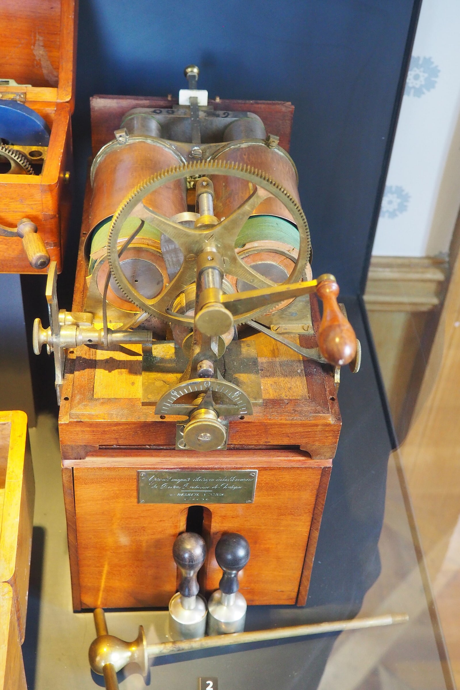
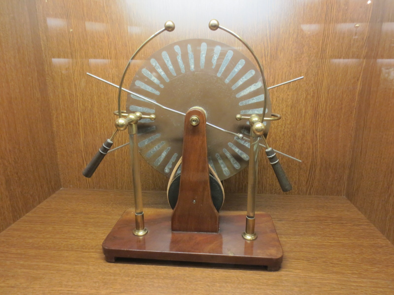
Portable “medical batteries” are the most common find — fitted wooden cases (mahogany or walnut) holding an induction coil, wet or dry cells, a vibrating make-and-break interrupter, and electrode handles with sponge or brush tips. A silk-lined case, exposed hand-made brasswork, and wet-cell fittings point earlier (1870s–1890s); dry cells, moulded bakelite fittings, and mains-adapter provision point later (1900s–1920s). Induction (Ruhmkorff-type) coils — two concentric windings, the secondary far finer — most commonly date to around 1900. Static and influence machines — Wimshurst, Holtz, Toepler-Holtz — are identifiable by their twin counter-rotating varnished-glass discs, tinfoil sectors, metal collecting combs, and Leyden-jar condensers (see Static Electricity Machines). Commercial Wimshurst machines by firms such as Baird & Tatlock were built roughly between 1890 and 1920. Glass “shock boxes” and coin-operated grip-testers are the fairground descendants of the same faradic coil and are frequently misidentified as purely medical.
5.7 Makers’, Patent, and Retail Marks
The surest dating evidence is on the object itself. Look for engraved or gilt maker’s plaques on the case lid, gold lettering on leather covers, patent-date stampings, and retailer labels inside the lid. American examples include Grigg’s Conical Electro Magnetic Machine, plaques of which cite a patent of 8 January 1873, and W.C. & J. Neff of Philadelphia, whose “Electro Magnetic Machines” carried gold lettering on leather. A stamped patent date is a terminus post quem — the machine cannot be older, though it may be considerably newer, as popular designs were made for decades. Because so many small makers competed and marks are often worn, the specialist reference for American apparatus is Dean P. Currier’s collector guide, Guide to Electrotherapy Instruments and History of Their American Makers (Dean P. Currier, 2004). Cross-reference any maker’s plate against museum object records (Science Museum Group, Smithsonian NMAH, the Bakken) before committing to a date.
5.8 The Real Hazards — and Why You Do Not Power Them Up
This guidance is strictly about not being harmed by an antique, and never about therapeutic use. These objects are safest treated as static museum pieces, cleaned and displayed but not operated.
- Static and influence machines are built to generate genuinely dangerous potentials. A working Wimshurst can reach roughly 50,000–60,000 volts; the Bakken lets visitors draw a spark from one for exactly that “wow” reason. A charged Leyden jar holds its charge and can deliver a painful, potentially harmful discharge even after the machine has stopped. Discharge and short jars before handling, and treat any influence machine as live.
- Cloth-wired mains coils and shock boxes were built long before earthing standards, modern insulation, or fuses. Perished rubber or cloth insulation, no ground, and a step-up secondary are a genuine electrocution and fire risk. Never plug in a vintage mains-powered unit “just to see.”
- Mercury interrupters, fitted to some induction coils (especially those repurposed for early X-ray work), contain liquid mercury — a toxic-vapour and contamination hazard if the reservoir is cracked or spilled. Handle suspect units in a ventilated space and do not disturb the mercury.
- Early X-ray and high-voltage adjacency. After 1896 the powerful Wimshurst became a high-voltage source for early Röntgen-ray experiments — and turn-of-the-century X-ray apparatus delivered on the order of a thousand times the dose of a modern machine (one study of surviving apparatus measured roughly 1,500×). Any antique associated with early radiography should be treated as a potential radiation as well as electrical hazard and, if in doubt, assessed by a specialist. The associated radium and radioactive “cures” of the era (see the Quack wing) carry their own real contamination risk.
The single rule that covers all of these: identify and display, do not energise.
5.9 Where They Live: Museums, Ethics, and Display
The serious collections set the standard for both identification and context. The Bakken Museum in Minneapolis is the world’s only museum devoted to medical electricity, holding roughly 2,000 scientific instruments and some 11,000 written works — electrostatic and magneto-electric generators, induction coils, and physiological instruments — and it is the reference point of first resort for this field. The Science Museum Group and Wellcome Collection in Britain, and the Smithsonian’s National Museum of American History, hold deep electrotherapy and physiotherapy holdings with online object records that are invaluable for dating a specimen against a documented example.
Displaying these objects well means telling the truth about them: crediting the real electrophysiology they helped establish, and marking plainly where the marketing outran the medicine. A cased violet-ray set is a legitimate high-frequency device and a cure-all fraud, and honest labelling says both. The ethical line for a private collector mirrors the museum’s: present the history, never imply the device is safe to use or that its therapeutic claims were true, and handle the genuine hazards — high voltage, mercury, early-radiography adjacency — soberly rather than as spectacle. Do that, and an antique medical battery becomes what it should be: a well-documented milestone on the road from amber and the Leyden jar to the defibrillator and the pacemaker.
Sources
- Ball & Featherstone, “Early history of defibrillation,” Anaesthesia and Intensive Care (2019)
- “On Occasion of Seventy-five Years of Cardiac Defibrillation in Humans,” PubMed (2023)
- Resuscitation Central — History and Science of Defibrillation
- Engineering and Technology History Wiki — Pacemakers
- Smithsonian NMAH — Pacemaker (object record)
- Johnson, “Transcutaneous electrical nerve stimulation (TENS)” (chapter PDF)
- “Resolving Long-Standing Uncertainty about the Clinical Efficacy of TENS,” PMC
- MedLink Neurology — Overview of electromyography and nerve conduction studies
- “History of electromyography and nerve conduction studies: A tribute to the founding fathers,” PubMed
- “The history of electrically-assisted transdermal drug delivery (iontophoresis),” PubMed
- Rehab Management — Therapeutic Electricity
- The Bakken Museum
- “The Bakken Library and Museum Artifacts Now Accessible Online,” PMC
- History of Science Museum, Oxford — Wimshurst machine
- History of Science Museum, Oxford — Wimshurst twin-plate machine and X-ray material
- Museum of Radiation and Radioactivity (ORAU) — Wimshurst Static Machine
- Science Museum Group — Wimshurst Electrical Machine (object record)
- “The Medical Battery in the United States (1870–1920),” ResearchGate
- ABC News — 1896 X-ray machine radiation compared with modern doses
Comments (0)